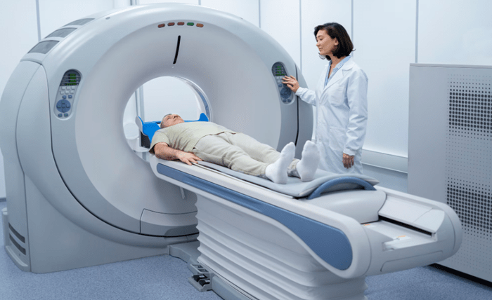
Medical imaging technology has evolved since Wilhelm Roentgen first captured bones on photographic film. Today’s digital medical imaging goes beyond fractures, delivering detailed cross sections, enhanced soft tissue contrast and real time motion analysis. This shift in digital scans in healthcare also brings AI driven insights and faster results without the chemical processing or long wait times of the past.
Whether you are a radiology professional, a healthcare leader or simply curious about diagnostic imaging advancements and the future of medical imaging, this article will guide you through:
- The evolution of imaging from first X-rays to modern CT and MRI systems
- How digital detectors and networked workflows speed diagnoses and reduce radiation doses
- Advanced techniques like 3D and 4D reconstructions and AI powered analysis
- Clinical use cases ranging from burn assessment to surgical planning
- Challenges and medical imaging trends that will shape the next generation of scans
By the end, you will understand how digital transformation in imaging technology improves diagnostic accuracy, cuts costs and supports personalized patient care. Let us begin by tracing the early breakthroughs that paved the way for today’s digital revolution.
Evolution of Medical Imaging Modalities
Roentgen’s X-Ray Discovery
In 1895 Wilhelm Conrad Roentgen observed a new type of ray using a Crookes tube. His first radiograph of his wife’s hand showed bones and some soft tissue contrast. Within months, X-ray labs appeared worldwide. By 1901, Roentgen won the Nobel Prize in Physics, launching a wave of medical imaging innovation.
Early Tomography and Its Limits
Conventional tomography emerged in the early 20th century to isolate a single anatomical plane. By moving the X-ray source and detector, clinicians could focus on specific structures. Soft tissue detail remained limited, and the method covered only small regions. These constraints set the stage for diagnostic imaging advancements in cross sectional imaging.
Birth of Computed Tomography
In 1967 Sir Godfrey Hounsfield introduced the first CT scanner. On October 1, 1971, the first patient underwent a CT scan that produced cross sectional images. This method revealed soft tissue contrast unseen on X-rays. Annual CT studies grew from a few thousand to over 100 million today, marking a milestone in medical imaging trends.
Advent of Magnetic Resonance Imaging
Following Nobel winning nuclear magnetic resonance research in the 1950s, Paul Lauterbur produced the first MRI images in 1973. MRI uses a static magnetic field and radio frequency pulses to excite hydrogen protons. As protons relax, they emit signals mapped into high contrast soft tissue images without ionizing radiation, reflecting a major step in medical imaging innovation.
Digital Transformation: From Film to Pixels
Medical imaging technology has shifted from chemical film processes to fully digital systems. This digital transformation replaces analog film-screen methods with flat panel detectors and computed radiography plates.
Electronic detectors capture images instantly, enabling faster, safer and more sustainable radiography. Digital medical imaging supports non-invasive imaging techniques and streamlines workflows across departments, making digital scans in healthcare more efficient and cost effective.
From Film-Screen to Digital Detectors
Early X-ray relied on photographic film and darkroom chemicals. Digital radiography uses flat panel detectors or computed radiography plates to capture images electronically. This shift eliminates film development time and physical storage needs. Hospitals can now archive images digitally in PACS, benefiting both clinicians and patients through rapid access and improved image quality.
Immediate Access and Workflow Efficiency
Digital images transmit instantly to workstations or a Picture Archiving and Communication System (PACS). Clinicians can view and adjust contrast, zoom with advanced software and share images across departments within seconds. Reduced wait times allow same-visit diagnoses, improving patient satisfaction and supporting data driven decision making in diagnostics.
Safety and Environmental Advantages
Switching to digital detectors can lower radiation dose by up to 90 percent compared to film-screen systems. It also removes hazardous chemical processing from the imaging chain, cutting medical waste and reducing a department’s environmental footprint. These improvements in environmental sustainability highlight a key benefit of medical imaging innovation.
Long-Term Cost Benefits
Although digital X-ray units require higher initial investment, hospitals save over time on film, chemicals and storage. Automated post-processing and streamlined workflows further boost departmental efficiency and return on investment. Digital medical imaging trends show that long-term cost savings outweigh upfront expenses.
Advanced Imaging Techniques Beyond X-Ray
Modern imaging extends beyond X-ray to include CT, MRI and ultrasound. These modalities enhance the sensitivity and specificity of anatomical and pathological assessment and represent key diagnostic imaging advancements.
Computed Tomography (CT)
Computed tomography uses rotating X-ray sources and advanced detectors to capture thin slices in fractions of a second. Modern scanners achieve submillimeter slice thickness and cut radiation dose by over fifty percent using iterative reconstruction algorithms. These improvements illustrate ongoing diagnostic imaging advancements in CT.
Key CT Applications
- Perfusion imaging for rapid detection of cerebral ischemia and penumbra
- Coronary CT angiography for noninvasive evaluation of coronary artery disease
- Spectral CT to enhance tissue contrast by capturing multiple photon energies
Magnetic Resonance Imaging (MRI)
MRI relies on nuclear magnetic resonance to excite hydrogen protons without ionizing radiation. Varying pulse sequences (T1, T2, diffusion) highlight different tissue properties for neurological, musculoskeletal and oncologic imaging. High field MRI systems deliver improved signal to noise ratio and spatial resolution, driving innovation in soft tissue diagnosis.
High-Field MRI Benefits
- 3.0 Tesla systems boost signal to noise and spatial resolution
- Wide bore designs improve patient comfort during longer scans
Ultrasound Imaging
Ultrasound employs high frequency sound waves for real time, radiation free visualization. Its portability and affordability make it ideal for point of care diagnostics, prenatal monitoring and interventional guidance. This non-invasive imaging technique supports rapid bedside evaluations and reduces the need for more complex scans.
Key Ultrasound Techniques
- Doppler imaging to assess vascular flow and velocity
- Elastography to evaluate tissue stiffness in soft tissue pathology
- Guided biopsies using live imaging for precise needle placement
Cutting-Edge Innovations: 3D/4D Visualization and AI Integration
Cutting edge medical imaging innovation leverages volumetric rendering and AI in medical imaging to expand clinical insights. 3D and 4D visualizations enable detailed anatomical models, while AI powered analysis accelerates image interpretation.
3D and 4D Volumetric Imaging
Three dimensional volume rendering creates detailed anatomical models from CT, MRI and ultrasound data. Clinicians can rotate and slice these reconstructions to examine structures from any angle. Wide area detectors like Canon Medical’s Aquilion ONE CT system capture dynamic volumes in millisecond intervals, supporting both anatomical and functional assessments.
3D Reconstructions in CT and MRI
Modern scanners use iterative reconstruction algorithms to reduce noise and improve resolution. Techniques such as breast tomosynthesis combine multiple X-rays into clear 3D mammograms, boosting lesion detection rates and exemplifying ongoing medical imaging innovation.
4D Dynamic Imaging
Four dimensional imaging adds time information to 3D datasets to track motion and physiology. In radiation oncology, 4D scans help map tumor movement and allow treatment planning that adapts to breathing cycles.
Applications include fetal assessment with real time 4D ultrasound for behavioral analysis, 4D MRI blood flow mapping in heart and vessels, and dynamic volume CT for joint motion in orthopaedic kinematics. These tools improve therapy accuracy and limit exposure to healthy tissue.
AI-Powered Image Interpretation
Emerging AI in medical imaging accelerates image analysis by automating segmentation and pattern recognition. Machine learning models can cut post processing times by up to 70 percent, uncovering details that manual review may miss. Integration of these tools into PACS and workstations supports data driven workflows.
Models such as U-Net and V-Net segment organs and detect anomalies, improving accuracy with large annotated datasets. AI powered systems flag critical findings and suggest measurements, boosting diagnostic speed and consistency while freeing radiologists for complex tasks in imaging technology in diagnostics.
Clinical Applications and Patient Benefits
Digital scans in healthcare deliver non-invasive insights that assist clinicians in assessment, planning and monitoring. High precision 3D data improves decision making and enhances patient outcomes.
Burn Assessment
High resolution 3D imaging maps burn wounds’ depth and surface area without direct contact. Clinicians can quantify burn severity to guide debridement and graft sizing, measure changes over time to adjust treatment plans and reduce the need for exploratory surgery. These non-invasive imaging techniques speed diagnosis and ensure tailored care from day one.
Surgical Planning
Advanced CT and MRI reconstructions enable virtual rehearsal of complex procedures. Surgeons benefit from personalized anatomical models for preoperative simulation, custom guides for implant alignment and reduced operating time with fewer complications. Collaboration with a dental crown lab allows precise design and fabrication of dental crowns and bridges.
Preoperative Simulation
Software platforms allow teams to simulate bone cuts or vessel anastomoses. This practice minimizes surprises in the operating room and boosts surgical confidence.
Wound Monitoring
Serial digital scans track healing by measuring volume reduction and tissue perfusion. Clinicians detect infection early through perfusion mapping, support remote follow up with objective metrics and engage patients with visual progress reports. By replacing subjective assessments, digital monitoring helps clinicians intervene sooner and improves overall satisfaction.
Challenges and Future Trends in Medical Imaging
As digital scans in healthcare generate massive datasets, hospitals face challenges in storage, computing and interoperability. Addressing these issues will drive the future of medical imaging.
Technical Hurdles
Modern scanners and AI tools produce large volumes of high resolution data that strain existing storage and compute resources. Hospitals must adopt scalable big data strategies, GPU accelerated pipelines and standardized annotation processes. Interoperability remains a hurdle due to heterogeneous data formats and legacy PACS systems, affecting seamless integration of medical imaging technology.
Ethical and Regulatory Barriers
AI driven analysis raises questions about patient privacy, informed consent and algorithmic bias. Providers must comply with HIPAA and GDPR requirements while ensuring transparency in model development. Medical devices that include machine learning face complex approval pathways, from FDA 510(k) clearances to EU MDR certifications under IEC 62304 software lifecycle standards.
Emerging Trends
Emerging medical imaging trends and signs of the future of medical imaging include:
- Cloud interoperability via DICOMweb and HL7 FHIR APIs for real time image streaming and vendor neutral archives
- Portable diagnostics such as handheld ultrasound probes and mobile CT scanners for remote or resource limited settings
- Integrative analytics combining imaging, genomics and electronic health record data for personalized medicine
Collaboration between clinicians, engineers and regulators will be key to overcoming current hurdles and unlocking the full potential of digital medical imaging.
Conclusion
Modern digital scans are reshaping medical imaging technology. Key takeaways include:
- Film processing has given way to electronic detectors, delivering instant images and reducing radiation exposure
- CT, MRI and ultrasound provide multi dimensional views with high contrast and zero chemical waste
- 3D reconstructions and 4D dynamic scans improve surgical planning, motion tracking and functional assessment
- AI in medical imaging automates segmentation and pattern recognition, speeding workflows and boosting consistency
- Clinical applications in burn assessment, surgical rehearsal and wound monitoring deliver clear patient benefits
- Managing large datasets, ensuring interoperability and meeting regulatory standards remain critical challenges
- Cloud archives, portable scanners and integrated imaging genomic analytics point to a more connected, personalized future
By embracing these innovations in digital medical imaging, care teams will diagnose faster, treat more accurately and drive better outcomes for years to come.

